Research Interests
Aggression is an essential social behavior across the animal kingdom
including humans. Since this behavior requires no learning, the underlying
neural circuit is believed to be hardwired in the brain. Attempts trying to
identify the brain substrates mediating aggression go back to 1920's. Early
electric stimulation and lesion studies in monkeys, cats, rats and hamsters
have identified the medial hypothalamus as critical to aggressive behavior,
but the fine anatomic structures remains unclear (Aggression, Violence and Brain, an interesting YouTube movie). Our current and future
research is aimed at understanding the aggression circuit in a genetically
tractable model system, mice. Genetic studies in mice have identified dozens
of genes which cause defects in aggression when disturbed, but the
functional studies in mice are largely lacking. Through gain and loss of
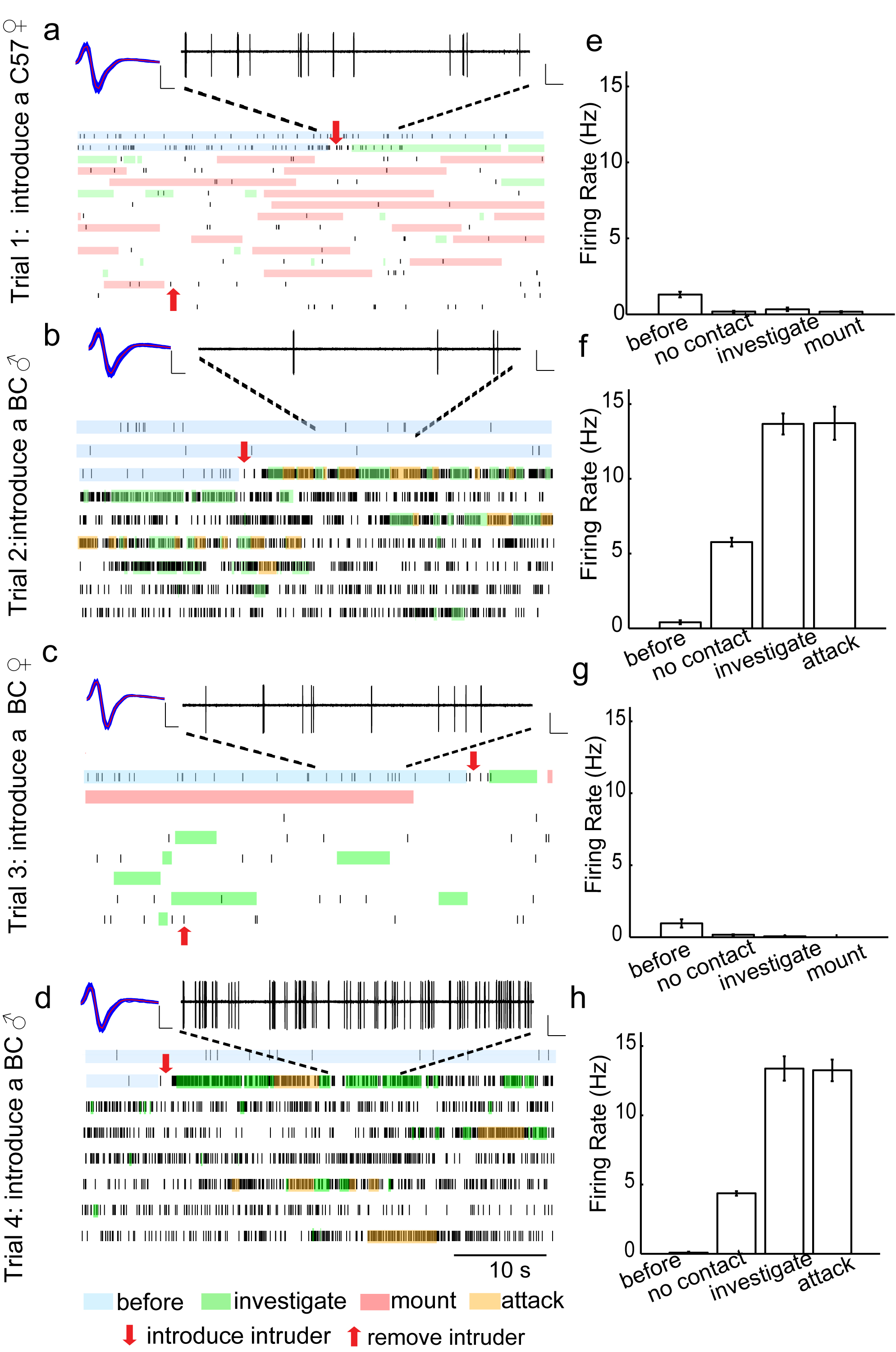
function manipulations and chronic recording in awake behaving mice (Figure 1), we have identified a small subnucleus in the hypothalamus, the
ventrolateral part of the ventromedial hypothalamus (VMHvl, Figure 2), as an essential aggression center in mice. The future study is aimed at expanding
our investigation on mouse aggression circuit from the VMHvl in multiple
directions.
Identify other relays in the mouse aggression circuit: Other relays in the
aggression circuit remain unclear. VMHvl forms an intricate network with
over 20 different brain regions. To understand how the sensory information
flows and transforms to elicit aggressive behaviors, we will systematically
manipulate the connections between the VMHvl and its upstream and downstream
targets and observe the behavioral change. We will further exam the cell activities in each potential relays using in vivo
chronic recording.
Aggression circuit in females: Social behaviors between males and females
differ in their motivation, execution and intensity. Even in human society,
the frequency of violence within females is much lower than that in males.
While electric stimulation of the similar region in the medial hypothalamus
in female rats is able to elicit aggression as in male rats, the VMHvl is
known to be sexually dimorphic and indispensable to the female sexual
behavior. It is thus interesting to understand whether the aggression
circuit in males and females is the same or not. If they indeed differ, is
the difference quantitative or qualitative?
Genetic dissection of the aggression circuit: The biggest advantage of
studying social behaviors in mice is the opportunity to relate genes to
behaviors. Many genes involved in various neurotransmitter neuropeptide and
hormonal systems are known to affect aggression but the exact mechanism is
unclear. With the knowledge of the important relays of the aggression
circuit, we can now exam the effect of the genetic manipulation on the
circuitry by in vivo recording in genetically modified animals. Furthermore,
combining optogenetics and recording, we could ask whether and how each
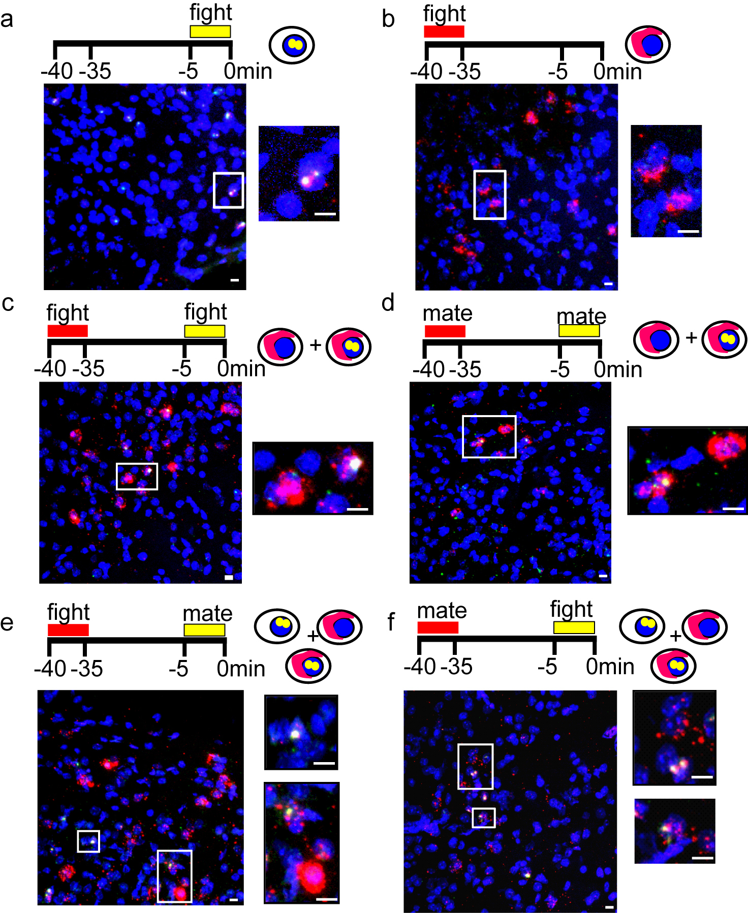
genetically defined population is involved in aggression. Interestingly, our
initial result indicates that aggression and reproduction circuits are
topographically intermingled and likely mutually inhibitory (Figure 3).
Genetically labeling and manipulating each population will allow us to
understand their interaction in further details.